|
|
| Home |
|
Prevention & Treatment of Cancers Research Projects: |
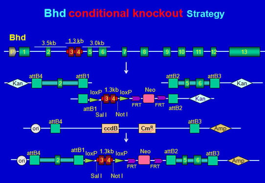
Birt–Hogg–Dubé (BHD) syndromeBirt–Hogg–Dubé (BHD) syndrome is an autosomal inherited cancer symdrome, first discribed by Birt, Hogg, and Dubé in 1977, characterized by the development of lesions/neoplasms in multiple organs. The featured lesions include skin tumors (hair follicle hamartomas/fibrofolliculoma, trichodiscomas, acrochordons), kidney tumors (34%chromophobe, 5% oncocytoma, 50% chromophobe/oncocytic hybrid, 9% CCRCC, 2% PRCC), spotaneous pneumothorax, and lung cysts. It is also associated with intestinal polyposis/colonic carcinoma, multiple lipomas, angiolipomas, parathyroid adenomas, and thyroid cancer/medullary carcinoma of the thyroid. As one of the hamartoma syndromes, BHD shares many clinical features (such as follicular hamartomas, mucosal fibromas, and internal malignancy) with Cowden syndrome (CD, affected gene PTEN), Peutz-Jeghers syndrome (PJS, affected gene LKB1), and tuberous sclerosis complex (affected genes TSC1/TSC2). Of these, Cowden syndrome shares the most clinical features with BHD, which implys that the gene responsible for BHD syndrome has similar roles. The BHD gene, located on chromosome 17p11.2, contains 14 exons spanning approximately 20 kb of genomic DNA and encodes a protein of 579 amino acids, folliculin (FLCN) that has no known functional domains. Germ-line mutations, somatic alterations, and loss of BHD mRNA have been observed in patients with BHD, colorectal cancer, and in some cases of gastric cancer. Germ-line mutations of the counterpart BHD have also been identified in dogs and rats having renal cell carcinomas and renal multiple cysts. Thus, BHD may be viewed as an important universal tumor suppressor gene.
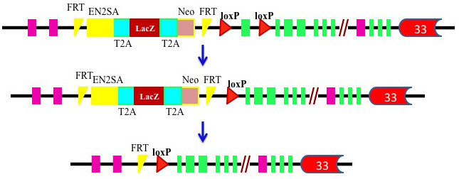
Since no in vitro experiments or nonmammalian model can replicate the complex processes of tumorigenesis in humans, the development of BHD-deficient animal models will shed light on the role of BHD in vivo and on the BHD-related biochemical pathways responsible for neoplasia, which eventually could lead to the development of therapeutic agents against BHD-related diseases. Although natural mutants could be used for experimental models, the possibilities of homozygous embryonic lethality and additional unknown genetic changes often impede further analysis of the phenotypes and the physiological role of the gene. The genetically engineered conditional knockout mouse model can bypass this barrier and provide a “cleaner” and more versatile system for functional studies of BHD gene protein FLCN. While it might be a suppressor of mouse cystogenesis demonstrated by a recent study, BHD is expected to be a potential tumor suppressor gene whose mutations have led to renal tumors and other diseases in BHD patients. Therefore, it is essential to further elucidate whether kidney-specific knockout of BHD in the mouse is also implicated in kidney tumorigenesis, and what mechanism is involved. Genetically engineered mouse models have become one of the most useful scientific tools in helping to understand the disease-related genes, particularly those that can only be examined in the context of an in vivo microenvironment. Development of an efficient Bhd-disruption mouse model will shed light on the pathologenesis and etiology of BHD syndrome and provide invaluable tools for testing new approaches to diagnosing and treating the diseases. By combining Gateway Technology with the Cre-loxP site-specific recombination system, we developed two kidney-specific Bhd knockout mouse models, a distal tubule/collecting duct–specific model (Bhd f/f-Ksp-Cre) and a proximal tubule–specific model (Bhd f/f-Sglt2-Cre).
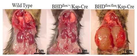
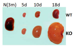
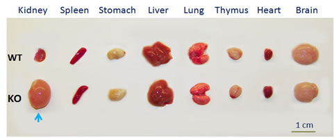
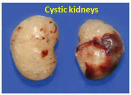
1. Distal tubule/collecting duct–specific mouse model (Bhdf/f-Ksp-Cre)Bhd f/f-Ksp-Cre (knockout, KO) mice developed enlarged kidneys characterized by polycystic kidneys, hyperplasia, and cystic renal cell carcinoma. The cystic kidneys in the affected BHDf/f-Ksp-Cre (KO) mice grow fast and reach up to 10 times larger than kidneys in wild-type (WT) mice within three weeks. The affected BHDf/f-Ksp-Cre mice died of renal failure at approximate three weeks of age, having blood urea nitrogen levels over tenfold higher than those of Bhd f/+-Ksp-Creand wild-type littermate controls. The other organs in the BHDf/f-Ksp-Cre (KO) mice appear normal, indicating the knockout is kidney-specific.
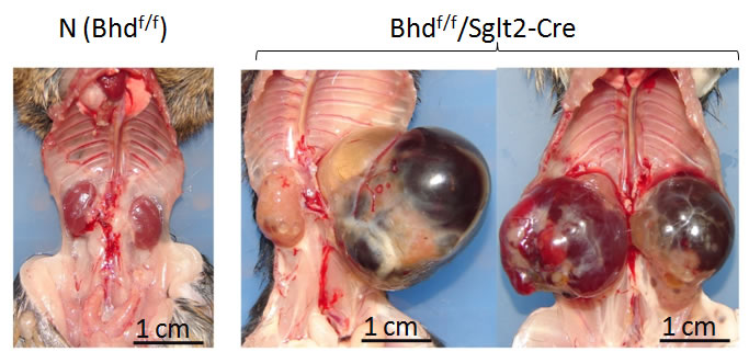
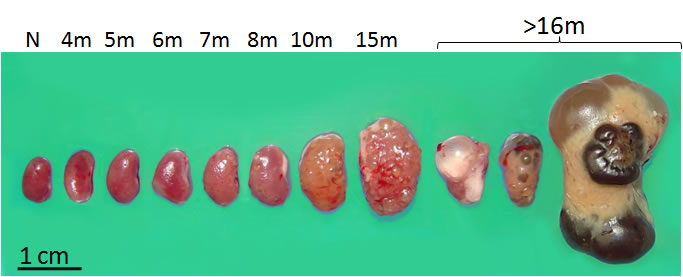
2. Proximal tubule–specific mouse model (Bhdf/f-Sglt2-Cre)The BHDf/f-Ksp-Cre mice rapidly developed polycystic kidney disease after birth and died in less than three weeks, probably due to disruption of Bhd in multiple types of kidney tubules. To obtain a kidney tissue-specific knockout mouse model having an extended lifespan, We generated a kidney-specific knockout mouse model by selectively disrupting the Bhd gene in the kidney proximal tubules through a Sglt2-Cre-loxP site-specific recombination system (Bhd flox/flox/Sglt2-Cre). The kidney proximal tubule is the transporter of filtered ions and solutes in the kidney, and most kidney cancers, especially clear cell RCCs and papillary RCCs, are hypothesized to arise from these tubules. In addition, our previous study demonstrated that Bhd is predominantly expressed in proximal tubules, implying that it has an important function in this region of the kidney. In addition, since the disruption of Bhd would be restricted to the proximal tubules, it is possible that affected mice would have a longer lifespan than Bhd flox/flox/Ksp-Cre mice and a higher potential to develop kidney tumors. Actually, unlike existing models, this proximal tubule–specific Bhd knockout mouse model developed multiple histological subtypes of renal neoplasms; in particular, the renal neoplasia appeared in a time-dependent order, presenting the progression of kidney tumorigenesis from hyperplasia to high-grade renal tumors, implying that kidney tumors, particularly clear cell and papillary RCCs, can originate from the kidney proximal tubule.
References: 1. Deficiency of FLCN in Mouse Kidney Led to Development of Polycystic Kidneys and Renal Neoplasia 2. Targeted disruption of Flcn in mouse kidney proximal tubule leads to development of renal cysts and multiple renal cancer histologies. |
